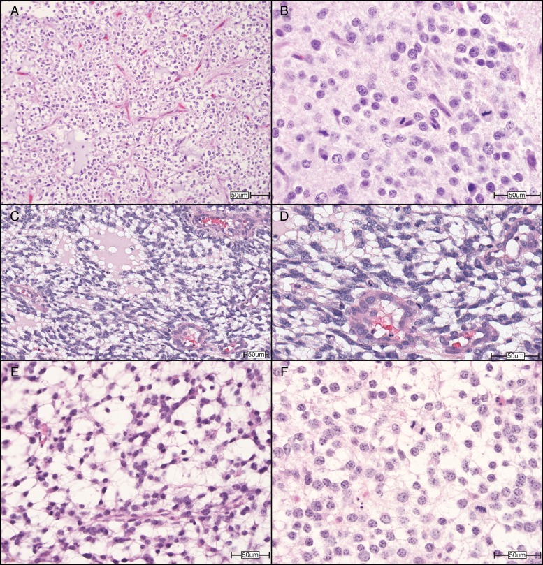
Fig. 3.
Representative photomicrographs of high-grade (WHO grade III) cerebral oligodendrogliomas in humans, dogs, and mice. (A) Human high-grade oligodendroglioma contains a delicate “chicken wire” vasculature and mucin-rich microcystic spaces and is composed of neoplastic cells with oval, hyperchromatic nuclei, and clear cytoplasm. (B) Higher magnification of a human high-grade oligodendroglioma shows tumor nuclei with crisp nuclear borders and abundant mitoses. (C) A surgical biopsy of a high-grade (grade III) cerebral oligodendroglioma in a Boston Terrier, demonstrating clear cytoplasm and distinct cell membranes, with ovoid hyperchromatic nuclei. (D) Higher magnification highlights endothelial hyperplasia of this lesion. (E) A high-grade oligodendroglioma allograft from a mouse stereotactically injected with oligodendrocyte progenitor cells harboring deletion mutations in transformation related protein 53 and neurofibromatosis type 1 (Ref PMID 25246577) shows similar features, including (F) the delicate vasculature and microcystic spaces and nuclei with crisp nuclear membranes and abundant mitoses. All sections were stained with hematoxylin and eosin. Original magnifications: 200× (A), 600× (B), 200× (C), 400× (D), 400× (E), 600× (F). Scale bars = 50 µm. Human and murine images courtesy of Dr C. Ryan Miller, University of North Carolina School of Medicine. Canine images courtesy of Dr R. Timothy Bentley, Purdue University College of Veterinary Medicine.

