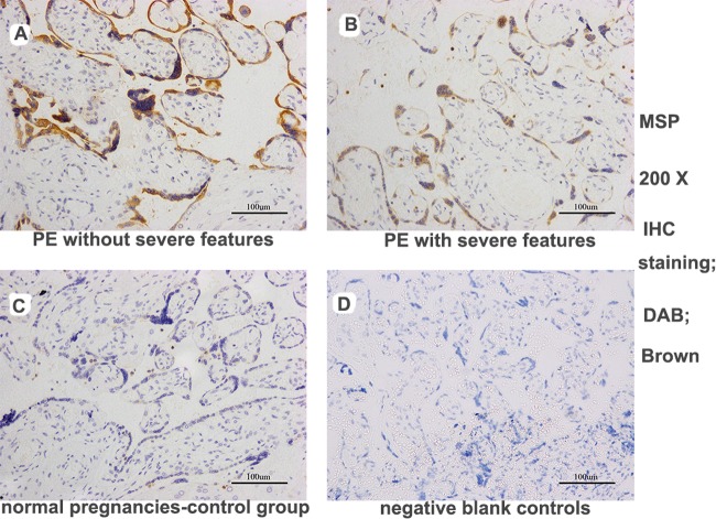
Fig 6. MSP expression in trophoblast cells in placental sections from pregnant women, as determined by immunohistochemical staining.
The PE group exhibited strong MSP staining in the placenta compared with the control group. Furthermore, increased MSP staining was detected in the sections from the patients without severe features vs. those with severe features in the PE group (DAB, brown: images A-D, 200X magnification). (images A: PE without severe features; images B: PE with severe features; images C: normal pregnancies in the control group; images D: negative blank controls).

