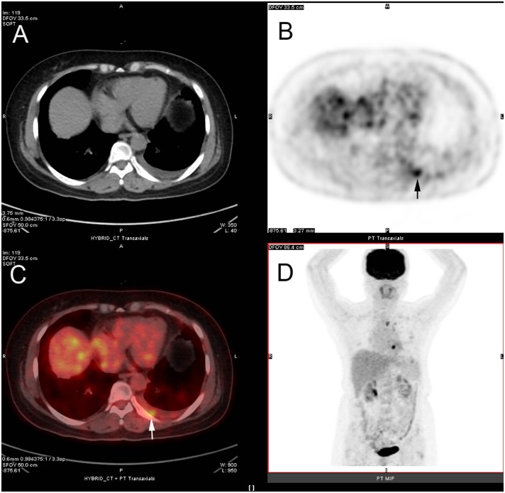
Fig 2. 18F-FDG PET/CT integrated imaging of 54-year old woman with left lung cancer and malignant pleural effusion.
Axial CT (A) shows effusion in left pleural cavity, and axial 18F-FDG PET (B, arrow) and axial fused 18F-FDG PET/CT (C, arrow) display nodular 18F-FDG uptake (SUVmax of 3.0) in left-posterior pleural region. Pathology from thoracentesis confirmed malignant pleural effusion caused by metastatic adenocarcinoma.

