
Figure 1.
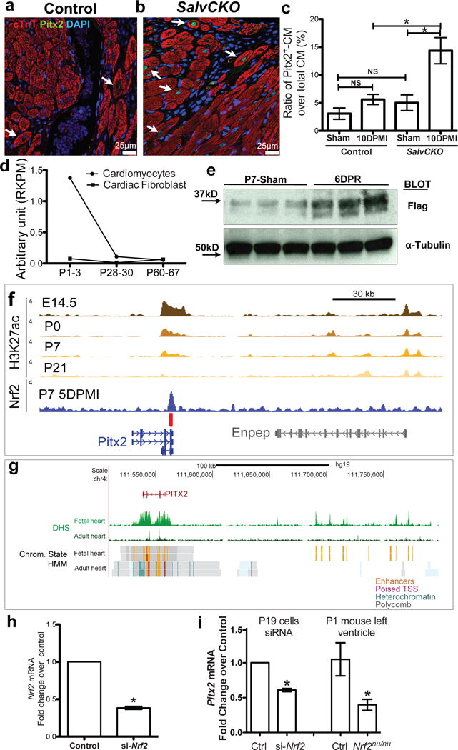
Pitx2 is induced in injured myocardial. (a–c) Border zone of SalvCKO (b) and control (a) hearts stained for Pitx2 (green), cTnT (red), and DAPI (blue) at 10 day-post-MI, with Pitx2+ cardiomyocyte ratio quantified in c, n=4. (d) Pitx2 expression showed by RNA-Seq, P, postnatal day. (e) Western blot of Flag and a-Tubulin in 5 DPR Pitx2flag ventricles, resected at P1. (f) Nrf2 directly binds to Pitx2 enhancer after LAD-O. The heart specific enhancers are marked by H3K27ac ChIP-Seq. red bar, Nrf2 binding element. (g) DHS-Seq and chromatin state tracks of fetal and adult human heart tissue. Orange color indicates active enhancer regions. (h) qPCR showed knocking-down of Nrf2 by siRNA in P19 cells, n=4. (i) qPCR of Pitx2 in P19 cells with siRNA targeting Nrf2, and Nrf2nu/nu heart, compared to controls, n=4. Mean ± S.E.M.; Statistical test, (c) one-way ANOVA plus Bonferroni post-test; (i, right part) Mann-Whitney; (h, i left part) see Methods; *, p<0.05; NS, not significant.