
Figure 2.
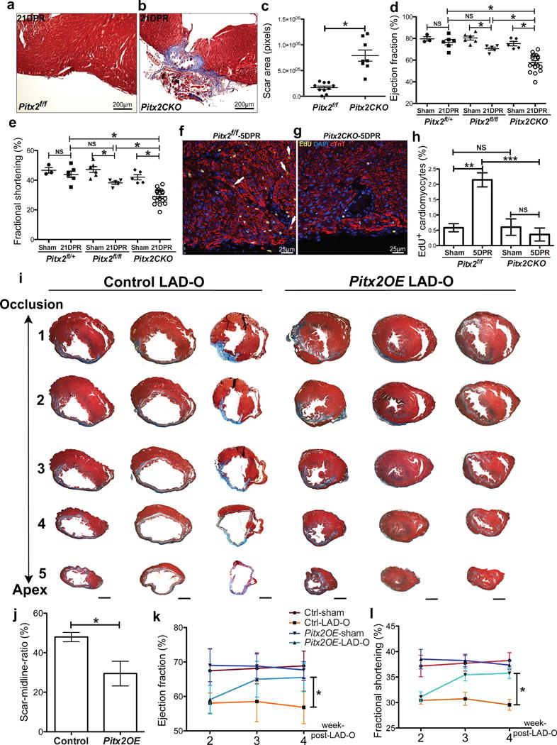
Pitx2 is required and sufficient to promote myocardial regeneration. (a–c) Trichrome-stained Pitx2f/f (a) and Pitx2CKO (b) apex at 21 DPR, with scar size quantified in c. (d, e) Echocardiography showed the ejection fraction (d) and fractional shorting (e) at 21 DPR. (f–h) 5 DPR Pitx2f/f (f) and Pitx2CKO (g) apical sections stained for EdU (yellow), cTnT (red), and DAPI (blue). Arrow, EdU+ cardiomyocyte. Cardiomyocyte proliferative ratio was quantified in h, n=4. (i) Serial transverse heart sections at 5 weeks post-LAD-O, performed at 8weeks. (j) Percentage of fibrotic left ventricular myocardium quantified at 5 weeks post-LAD-O, n=5. Scale bar, 1mm. (k, l) Ejection fraction (k) and fractional shortening (l) of LAD-O and sham hearts. Mean ± S.E.M.; Statistical test, (d, e) one-way ANOVA plus Bonferroni post-test; (c, h, j–l) Mann-Whitney; *, p<0.05; NS, not significant.