
Extended Data Figure 2.
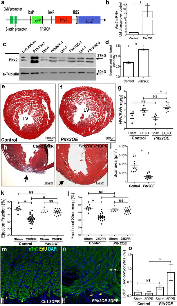
Pitx2 promotes myocardial regeneration after apex resection at P8. (a) Schematic of Pitx2-expressing construct (Pitx2Gof). (b–d) Pitx2Gof was crossed with Mhccre-Ert strain to generate Mhccre-Ert/+;pitx2Gof (Pitx2OE), after Tamoxifen treatment, qPCR (b, n=4), western (c, d, n=3) showed the over-expression of Pitx2 in Pitx2OE ventricles. (e–f) Trichrome-stained cross sections from 13 weeks old sham hearts of control (e) and Pitx2OE (f), with tamoxifen administrated at 7–8 weeks old. (g) Heart weight over body weight ratio of adult sham and LAD-O hearts. (h–j) Apex resection of Pitx2OE (i) and control (Mhccre-Ert/+) (h) hearts at P8 followed by trichrome staining at 28 DPR, scar area was quantified in j. (k, l) Echocardiography showed ejection fraction (k) and fractional shortening (l) at 28 DPR. (m–o) EdU labeling of Pitx2OE (n) and control (m) apical area, 8 days after P8 resection, sections were stained for cTnT (green), EdU (yellow), and DAPI (blue). Arrow indicates EdU-labeled cardiomyocytes, with quantification in o, n=4. Mean ± S.E.M.; Statistical test, (g, k, l) one-way ANOVA plus Bonferroni post-test; (b, d, j, o) Mann-Whitney. *, p<0.05; NS, not significant.