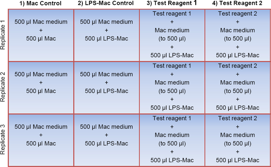
Figure 1. Sample plate layout for the macrophage assay.
To prepare the 12-well assay plate, the test reagents of interest are first added to the appropriate wells in triplicate (shown here in columns 3 and 4). Macrophage (Mac) medium is then added to every well to a final volume of 500 µl followed by addition of 500 µl of the Mac cell suspension to each well in the first column (unstimulated Mac control). 500 µl of the LPS-stimulated macrophages (LPS-Mac) are transferred to each of the remaining 9 wells (columns 2, 3, and 4). Note that three of these wells do not contain a test reagent (shown here in column 2) and therefore serve as the LPS-stimulated macrophage control (LPS-Mac control). The final volume per well is 1.0 ml. For simplicity, wells containing a vehicle control for the test reagents have been omitted.

