Figure 3.

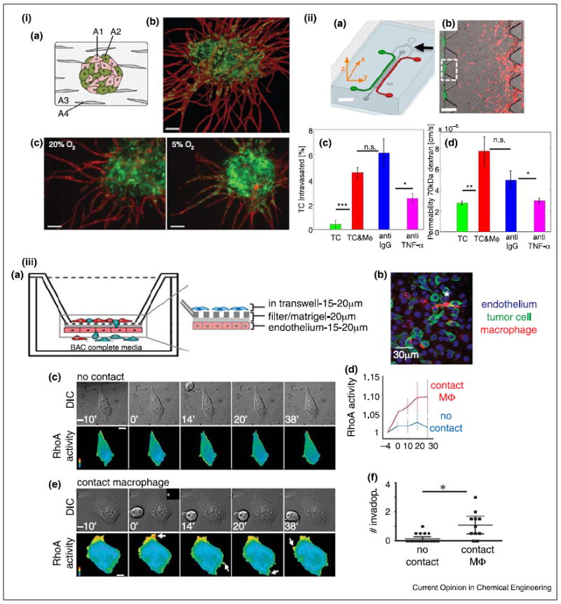
Models for tumor intravasation. (i) Prevascularized tumor (PVT) spheroid model, (a) Schematic of the PVT spheroid model. EC (A1) and tumor cell (A2) spheroids were co-cultured with fibroblasts (A4) in 3D fibrin matrix (A3), (b) PVT spheroid shows radial EC sprouting (CD31, red) from the prevascularized tumor (EGFP-transfected SW620, colon) spheroid. Scale bar is 100 μm. (c) Decreased oxygen tension increases intravasation of the SW620 cells. Scale bar is 100 μm. (ii) Microfluidic tumor-vascular interface model, (a) Schematic of the device. EC channel (green), tumor channel (red). Scale bar is 2 mm. (b) Fibrosarcoma cells (HT1080, red) invade through the ECM toward EC (green). Scale bar is 300 μm. (c) Macrophages enable tumor intravasation though TNF-α signaling, (d) Enhanced tumor intravasation is endothelial permeability dependent. (iii) Transwell in vitro model for tumor intravasation. (a) Schematic of the transendothelial migration of cancer cells in the presence of macrophages. (b) A representative image of apical section of the transwell, (c) RhoA activity in cancer cells in the absence of direct contact of macrophages, (d) RhoA activity in cancer cells with or without direct contact of macrophage, (e) RhoA activity in cancer calls in direct contact of macrophages, (f) Tumor invasion with or without direct contact with macrophage. Scale bars (a,b) are 10 μm. (i) Reproduced from [37••] with permission from the Royal Society of Chemistry; (ii) reproduced from [34] with permission from the National Academy of Sciences; (iii) reproduced from [36•] with permission from the Nature Publishing Group.
