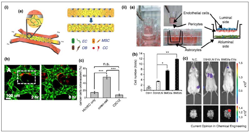
Figure 4.

Models for tumor extravasation. (i) Breast tumor extravasation in the bone-mimicking microenvironment (BMi). (a) Schematic of the microfluidic device. ECs, MSCs, and osteoblasts (OBs) were initially seeded in fibrin gel. After formation of vascularized BMi, cancer cells (CC) were added to the side channels. (b) Cancer cell (red) extravasation through HUVEC (green) network in the BMi. (c) Cancer cell extravasation enhanced by osteocells. Myoblasts, C2C12, were used as a control. (ii) Breast tumor extravasation in the brain microenvironment. (a) Schematic of the in vitro model of blood–brain barrier (BBB). (b) Tumor extravasation in the presence of extracellular vesicles (EVs) from different tumor cells (D3H1: primary tumor cells, D3H2LN: lymph node metastases, BMD2a and 2b: brain metastases). Brain metastases derived EVs enhanced breast tumor extravasation. (c) Bioluminescence images of D3H2LN and BMD2a derived EVs injected mice (negative control, N.C.). BMD2a-derived EVs promoted brain metastasis in vivo. (i) Reproduced from [47••] with permission from the National Academy of Sciences and (ii) reproduced from [46•] with permission from the Nature Publishing Group.
