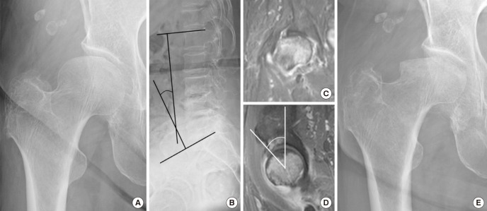
Fig. 2.
A 77-year-old woman with a subchondral insufficiency fracture of the right femoral head. (A) Radiograph after sudden onset of right hip pain shows subchondral collapse in the superolateral portion of the femoral head. (B) Lateral radiograph of the lumbar spine shows decreased lumbar lordosis (24.7°). (C) On the mid-coronal T2-weighted MRI, a diffuse bone marrow edema pattern with linear low signal intensity line at epiphysis of femoral head is seen. (D) On the mid-sagittal T2-weighted MRI, anterior center-edge angle was decreased to 29.2°. (E) Radiograph at 2 months follow-up shows rapidly progressive collapse of the femoral head.

