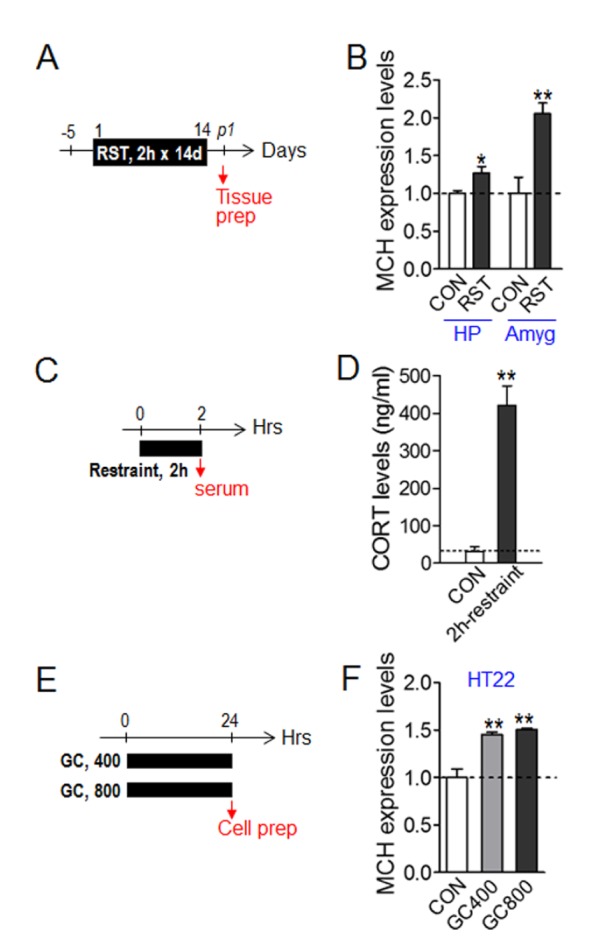
Fig. 3. Repeated restrant stress upregulated the expression of MCH in the brain and corticosterone increased the expression of MCH in HT22 cells. (A) Experimental design for treatment with 2 h×14 d RST, and the time point for brain tissue preparation (arrow). (B) Real-time PCR showing increased expression of MCH in the hippocampus (HP) and amygdala (Amyg) after treatment with repeated restrant stress (n=6 each). (C) Experimental design for treatment with a single 2-h restraint and the time point for blood preparation (arrow). (D) Corticosterone levels in the blood of mice treated with 2 h restraint (restraint) and their control (CON) (n=7 each). (E) Experimental design for treatment with corticosterone (final concentration: 400 or 800 µg/ml) and the time point for cell harvesting (arrow). (F) Real-time PCR showing increased expression of MCH in HT22 cells after treatment with corticosterone (n=5 animals each; three repeats of duplicated PCR data). Data are presented as mean ± SEM. * and **, p<0.05 and p<0.01, respectively for the differences between the control and indicated groups (one-way ANOVA and Newman-Keuls post hoc test).