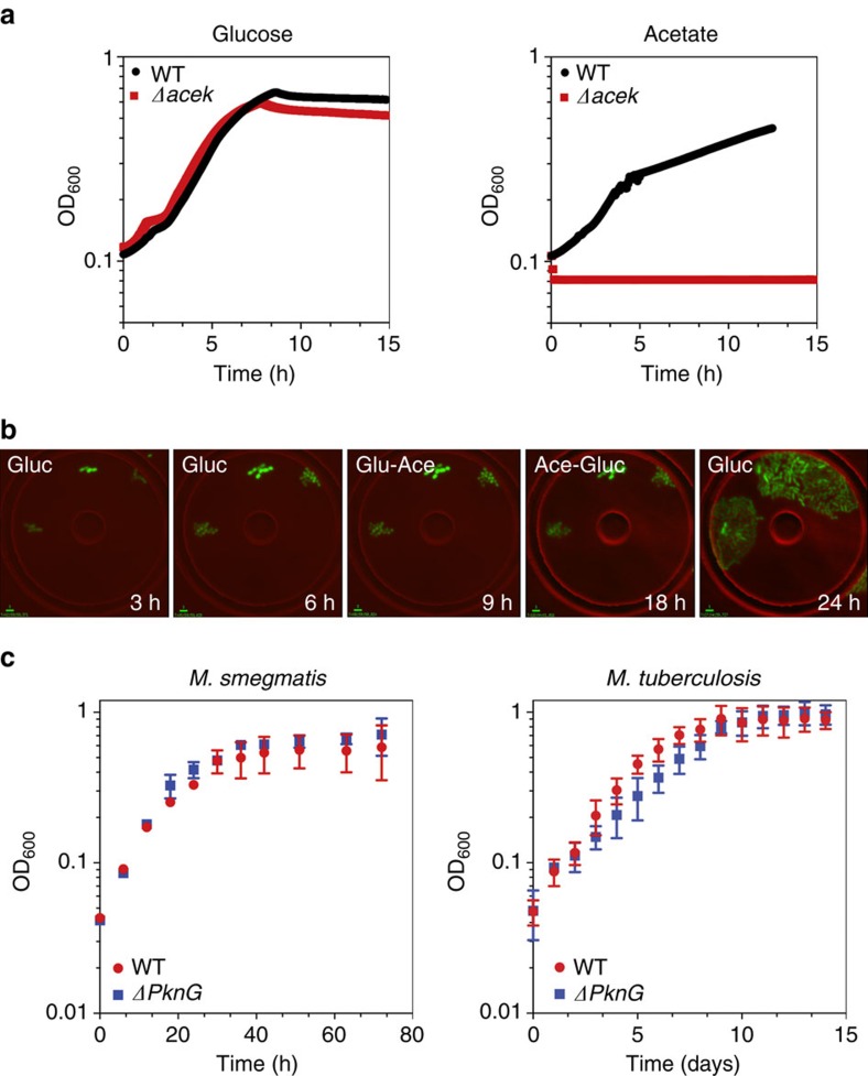
Figure 3. Phosphorylation-mediated branch-point regulation.
(a) Growth (OD600) of wild-type and ΔaceK strains of E. coli on minimal medium containing glucose (left) or acetate (right). Data are means±s.d. (n=3 independent experiments). (b) Time-lapse series of GFP-expressing E. coli ΔaceK cells grown in a microfluidic device and imaged on phase-contrast and fluorescence channels (merged) at 5-minute intervals. Flow medium was M9 minimal medium containing glucose (0–6 h) before switching to acetate (6–18 h) and then back to glucose (18–24 h). Scale bar, 3 μm. (c) Growth (OD600) of wild-type and ΔpknG strains of M. smegmatis (left) or M. tuberculosis (right) on minimal medium containing acetate. Data are means±s.d. (n=3 independent experiments).

