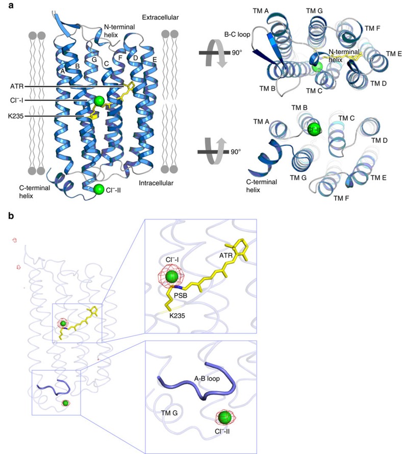
Figure 1. Crystal structure of the ClR.
(a) ClR structure in a ribbon model. Transmembrane helices A–G (TM A–TM G) are indicated and the putative membrane parts are drawn schematically. (b) Anomalous difference Fourier map of Br− shown as red meshes (contoured at 5.0σ). Magnified views of the peaks of the map are shown in boxes. Two chloride ions bound to ClR structure are depicted by green spheres. All-trans retinal (ATR), linked covalently to K235, is shown in a yellow stick model.

