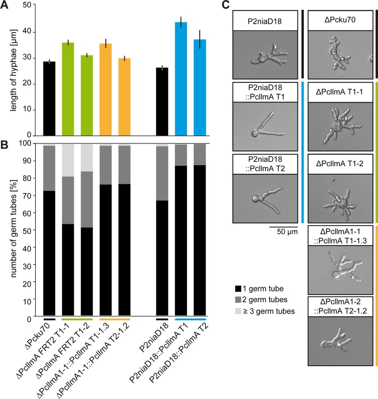
FIG 6 .
Hyphal morphology of germinating conidia. (A) Lengths of germinating hyphae were measured after 18 h of cultivation on solid CCM. Values are the mean scores of 300 independent measurements; averages ± standard deviations are indicated. (B) Numbers of germ tubes per germinating conidiospore were determined for 300 independent spores after 18 h of cultivation on solid CCM. Values are given as percentages of all analyzed hyphae per strain. Black, 1 germ tube; dark gray, 2 germ tubes; light gray, ≥3 germ tubes per conidiospore. (C) Representative micrographs of germinating conidiospores analyzed in panels A and B. Scale bar = 50 µm.

