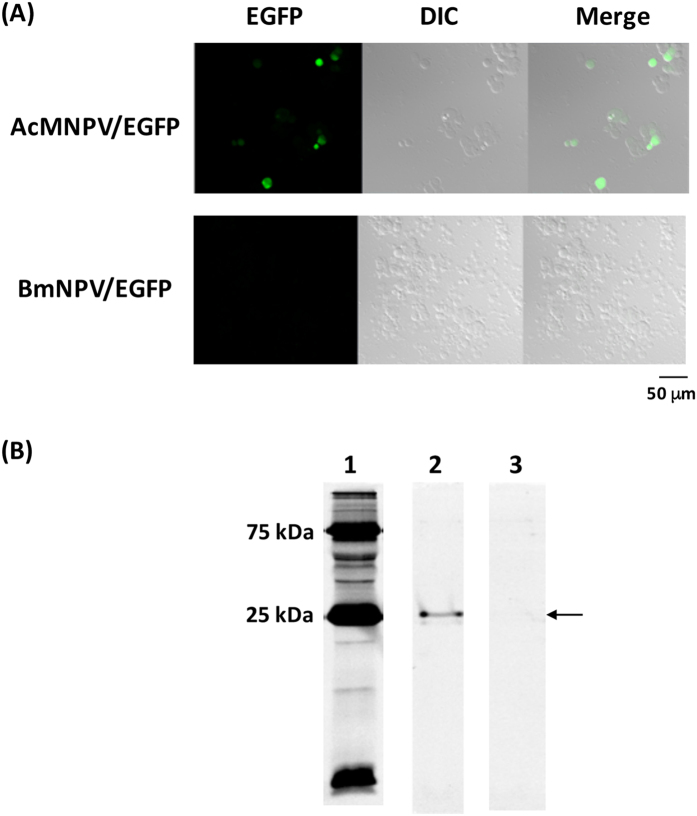
Figure 1. Transduction of AcMNPV/EGFP and BmNPV/EGFP into HEK293T cells.
(A) Fluorescence microscopy of HEK293T cells transduced with each recombinant baculovirus. Each baculovirus was transduced into mammalian cells at M.O.I. 300, followed by cultivation for 48 h. After 48 h cultivation, trypsinized cells were put onto a glass slide and green fluorescence in the cells was observed using confocal laser scanning microscopy. (B) SDS-PAGE of baculovirus-transduced HEK293T cell homogenates. Lane 1: Marker, Lane 2: AcMNPV/EGFP, Lane 3: BmNPV/EGFP. Precision plus protein dual color standard (Bio-Rad) was used as a protein marker. An arrow indicates expressed EGFP.

