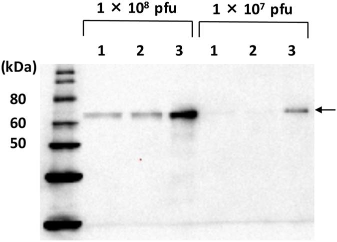
Figure 6. Western blot of GP64 from each baculovirus.

Each virus was propagated on Bm5 (BmNPVΔbgp/AcGP64/EGFP and BmNPVΔbgp/BmGP64-EGFP) or Sf-9 (BacMam 2.0) cells and partially purified. Subsequently, 1 × 10⁸ or 1 × 10⁷ PFU of each virus was separated by SDS-PAGE, transferred to a PVDF membrane, and subjected to western blot analysis using rabbit anti-BmNPV GP64 polyclonal antibody. Lane 1: BmNPVΔbgp/AcGP64/EGFP, Lane 2: BmNPVΔbgp/BmGP64-EGFP, Lane 3: BacMam 2.0. Arrows indicate expressed AcGP64 or BmGP64.
