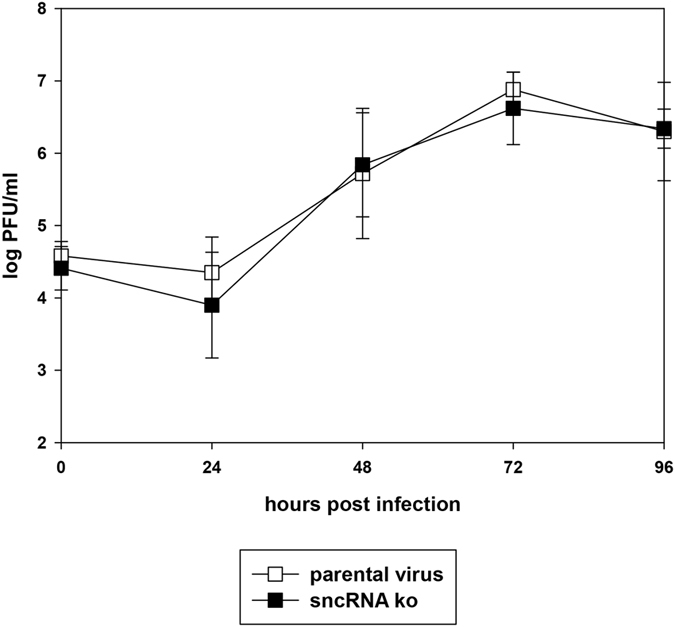
Figure 3. Lytic virus replication in vitro.

NIH3T3 cells were infected with the indicated viruses at an MOI of 0.1. Cells and cell culture supernatants were harvested at different time points after infection, and virus titers were determined by plaque assay on BHK-21 cells. Data shown are the means ± SD from three independent experiments.
