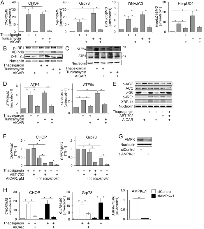
Figure 2. AICAR inhibits ER stress responses.
(A) mRNA expression of ER stress markers, (B,C) Western analysis of cell lysates (B) or nuclear extracts (C).(D) mRNA expression of ATF4 and ATF6α in macrophages pre-exposed for 1 h to AICAR and treated with thapsigargin or tunicamycin for 6 h. (E) Western analysis of macrophages pre-exposed for 1 h to 0.5 mM AICAR in the absence or presence of ABT-702 and treated with thapsigargin for 6 h. (F) mRNA expression of CHOP and Grp78 in macrophages pre-exposed for 1 h to indicated concentrations of AICAR in the absence or presence of ABT-702 and treated with thapsigargin or tunicamycin for 6 h. (G) Protein expression of AMPK 72 h post-transfection with control siRNA or AMPKα1 siRNA. (H) mRNA expression of CHOP, Grp78 and AMPKα1 in macrophages transfected with control siRNA or AMPKα1 siRNA for 72 h prior to treatments with thapsigargin for 6 h with or without 1 h AICAR pre-exposure. *p < 0.05. Data represent mean values ± SE of at least three independent experiments. ns, non-specific band. Western Blot images are cropped scans either of the same or of the duplicate membranes probed with different antibodies.

