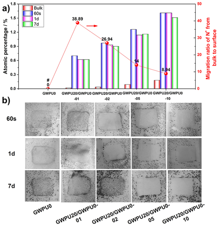
Figure 4. Surface structure and contact-active antibacterial activity of GWPU blends films.
(a) Atomic percentages of N+ on the surface of the GWPU blend films (after 60 s, 1 day, and 7 days of washing in water, respectively) and migration ratio of N+ from bulk to surface of GWPU blend films obtained from XPS spectra. # no N+ was detected in GWPU0. (b) Photographs of GWPU blend films coated on glass slides with the size of 1.5 cm × 1.5 cm (after 60 s, 1 day and 7 days of washing in water, respectively) and sprayed with S. aureus aqueous suspensions (106 CFU/ml) in phosphate buffer saline (PBS), air dried for 10 min, incubated with in a nutrient broth (0.8% agar) medium at 37 °C for 24 h, stained with 3 mL 5% 2, 3, 5-triphenyltetrazolium chloride (TTC). Each black dot corresponds to a bacterial colony grown from a single surviving bacteria cell.

