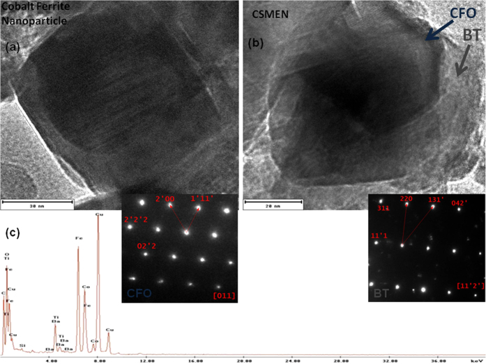
Figure 2.
Morphology and microstructure of the synthesized CSMENs demonstrated by Transmission Electron Microscopy image and the selective area diffraction patterns insets (a) for the CFO core and; (b) for the CSMEN’s BT shell; (c) The energy dispersive spectrum for the composition analysis of the CSMEN.

