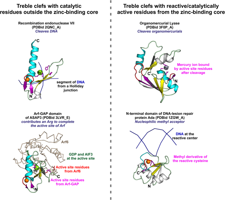
Figure 2. Comparison of all catalytic treble clefs.
The protein structures on the left are of catalytic TCs whose active site resides outside the zinc-binding core and those on right are of TCs whose reactive/active-site residues are from the zinc-binding core. The basic colouring scheme follows Fig. 1; side-chains of active-site/reactive residues are coloured magenta. TCs are shown as ribbons and the DNA/protein partners are shown as backbone trace.

