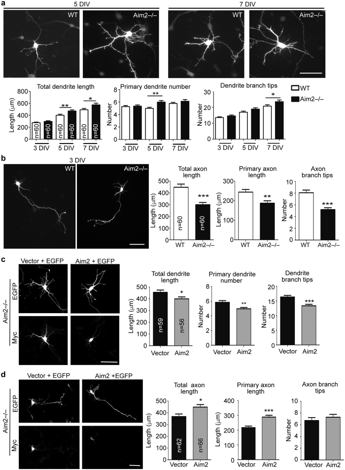
Figure 2. Deletion of Aim2 inhibits axonal growth but promotes dendritic growth.
(a,b) Cortical and hippocampal mixed cultures prepared from Aim2−/− and WT embryos were transfected with EGFP at 1 DIV and subjected to immunostaining using GFP and SMI-312 R (axonal marker) or MAP2 (dendritic marker) antibodies at different time points as indicted. After identifying neurite property (either axons or dendrites) based on SMI-312R or MAP2 signals, the lengths of axons and dendrites were determined by GFP signals. (a) Dendritic phenotypes. (b) Axonal phenotypes. (c,d) Aim2−/− cortical and hippocampal mixed cultures were transfected with Myc-tagged Aim2, EGFP and control vector, as indicated, at (c) 2 and (d) 1 DIV and harvested for immunostaining at (c) 5 and (d) 3 DIV. The phenotypes of (c) dendrites and (d) axons were quantified as indicated. In (a,b), only GFP images are shown; in (c,d), both GFP and Myc signals are shown. Scale bar, 50 μm. Neurons were collected from two independent experiments. The sample sizes (n) of examined neurons are indicated. The data represent the mean plus s.e.m. *p < 0.05; **p < 0.01; ***p < 0.001.

