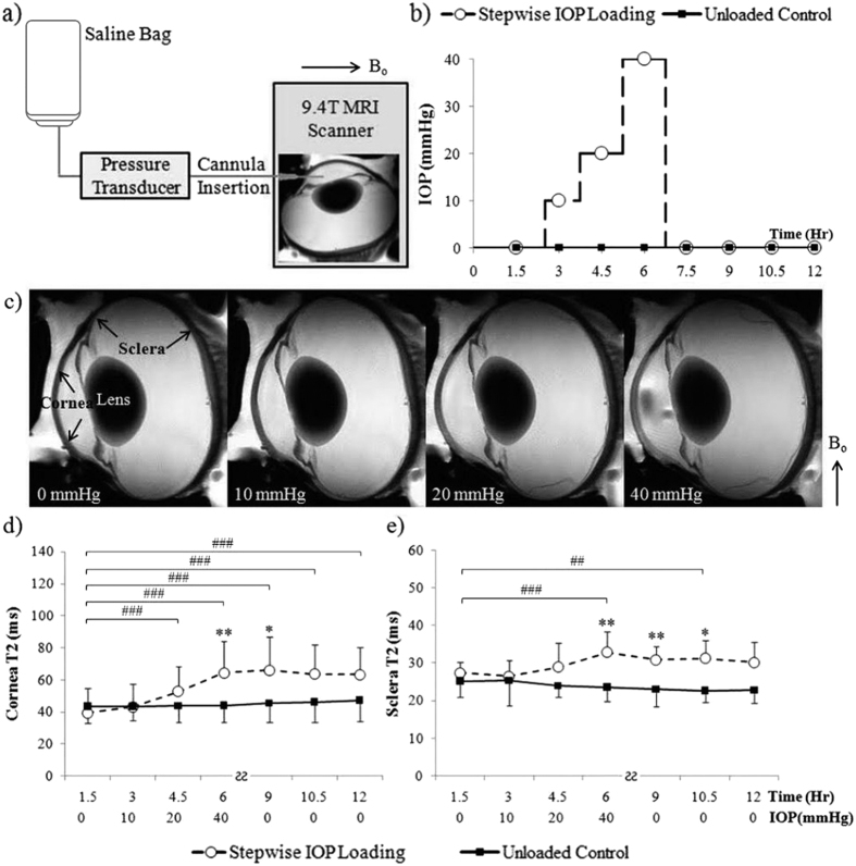
Figure 1. Dynamic imaging of the effects of stepwise intraocular pressure (IOP) changes on magnetic resonance relaxometry in the corneoscleral tissues of freshly prepared ovine eyes.
(a) Schematic diagram of anterior chamber perfusion to a fresh, unfixed ovine eye in the 9.4 Tesla MRI scanner. A plastic cannula was inserted into the anterior chamber and connected to a saline bag that was risen at different heights to induce different levels of IOP elevation. (b) Experimental paradigm of the stepwise IOP loading MRI experiment and the IOP-unloaded control experiment. (c) Representative ex vivo T2-weighted MRI (T2WI) of the same ovine eye loaded at 0, 10, 20 and 40 mmHg. (d,e) Quantitative comparisons (mean ± standard deviation) of transverse relaxation time (T2) in cornea (d) and sclera (e) upon stepwise IOP loading (dashed lines) and in unloaded control (solid lines). Both cornea and sclera T2 gradually increased as IOP increased from 0 to 40 mmHg (Tukey’s multiple comparisons tests between first MRI session and subsequent sessions, ##p < 0.01, ###p < 0.001; comparisons between other sessions are not shown here for clarity) but remained unchanged after being unpressurized (Tukey’s multiple comparisons tests between MRI sessions, p > 0.05). No significant change was observed in the unloaded control tissues over time (Tukey’s tests between MRI sessions, p > 0.05) (Sidak’s multiple comparison tests between IOP loaded and unloaded control tissues: *p < 0.05, **p < 0.01) (Bo: main magnetic field).

