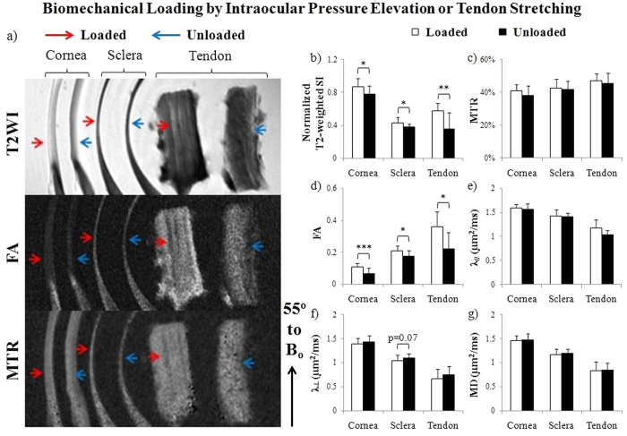
Figure 4. Microstructural organization and macromolecular contents of IOP-loaded sclera and cornea tissues, and stretch-loaded tendon tissues by T2-weighted MRI (T2WI), diffusion tensor MRI (DTI) and magnetization transfer MRI (MTI).
(a) Representative T2WI (top), fractional anisotropy (FA) maps (middle) and magnetization transfer ratio (MTR) maps (bottom) of loaded (red arrows) and unloaded (blue arrows) cornea, sclera and tendon tissue strips. The tissues were oriented and measured near the magic angle at ~55° to the main magnetic field (Bo) to enhance MRI signals for more sensitive examinations; (b–g) Quantitative comparisons (mean ± standard deviation) of (b) T2-weighted signal intensity (SI), (c) MTR, (d) FA, (e) axial diffusivity (λ//), (f) radial diffusivity (λ⊥) and (g) mean diffusivity (MD) between loaded and unloaded cornea, sclera and tendon strips. ANOVA tests showed significant differences between cornea, sclera and tendon tissues under the same loading conditions for each MRI parameter in (b–g) (p < 0.05) except loaded tissues in MTR (c) (p > 0.05) (Paired t-tests between loaded and unloaded cornea or sclera: *p < 0.05, **p < 0.01; ***p < 0.001; Unpaired t-tests between loaded and unloaded tendon: *p < 0.05).

