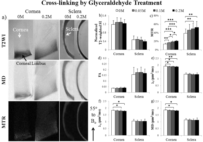
Figure 5. Microstructural and macromolecular changes in the fresh sclera and cornea tissues after glyceraldehyde cross-linking treatment with T2-weighted MRI (T2WI), diffusion tensor MRI (DTI) and magnetization transfer MRI (MTI).
(a) Representative T2WI (top), mean diffusivity (MD) maps (middle) and magnetization transfer ratio (MTR) maps (bottom) of cross-linked cornea (left panel) and sclera (right panel) after treating with 0.2 M glyceraldehyde solution or sham solution (0 M). The tissue strips were oriented and measured near the magic angle at ~55° to main magnetic field (Bo); (b–g) Quantitative comparisons (mean ± standard deviation) of (b) T2-weighted signal intensity (SI), (c) MTR, (d) fractional anisotropy (FA), (e) axial diffusivity (λ//), (f) radial diffusivity (λ⊥) and (g) MD in cornea and sclera strips after treatments with glyceraldehyde cross-linking solutions at 0, 0.05, 0.10 and 0.20 M concentrations. (Tukey’s multiple comparisons tests: *p < 0.05, **p < 0.01, ***p < 0.001).

