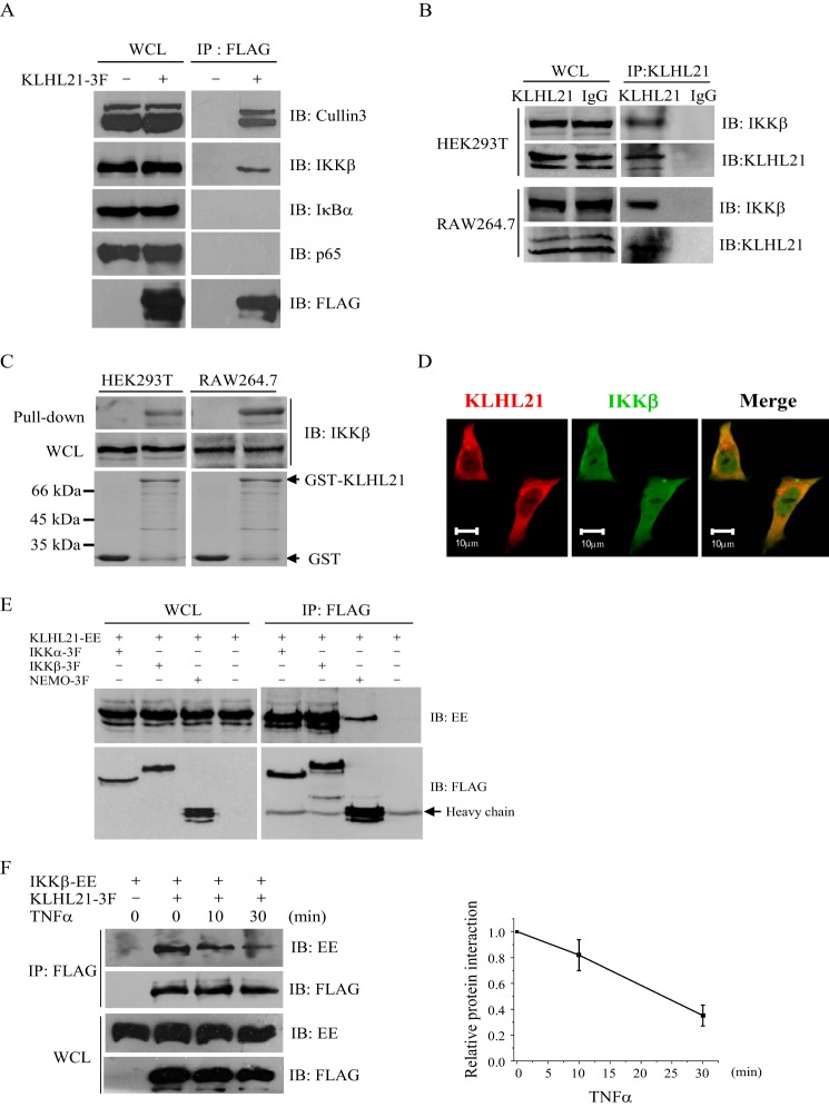
FIGURE 4.
KLHL21 protein specifically binds to IKKβ. A, 293T cells were transfected with KLHL21–3F. KLHL21-3F was immunoprecipitated with anti-FLAG beads and then analyzed by immunoblotting with the indicated antibodies. B, whole cell lysates prepared from 293T and RAW264.7 cells were immunoprecipitated with anti-KLHL21 polyclonal antibody and then analyzed by immunoblotting with the indicated antibodies. C, purified GST-KLHL21 fusion protein was incubated with whole cell lysates prepared from 293T and RAW264.7 cells, respectively. The precipitates after pull-down were analyzed by Western blotting with anti-KLHL21 antibody. D, HeLa cells were transfected with C-terminal mCherry-tagged KLHL21 (red). Cells were fixed and immunostained with anti-IKKβ antibody (green). E, 293T cells were cotransfected with vector expressing IKKα-3F, IKKβ-3F, NEMO-3F, and KLHL21-EE as indicated. Lysates were immunoprecipitated with anti-FLAG and then analyzed by immunoblot with anti-EE and anti-FLAG, respectively. F, 293T cells cotransfected with IKKβ-EE and KLHL21–3F were collected at the indicated time points after TNFα treatment. KLHL21-3F was immunoprecipitated with anti-FLAG beads and then analyzed by immunoblotting with the indicated antibodies. The density ratio of immunoprecipitated IKKβ/KLHL21 quantified by densitometric scanning is shown on the right (n = 3). All experiments were performed at least three times. IB, immunoblotting; IP, immunoprecipitation; WCL, whole cell lysates. Error bars, S.E.

