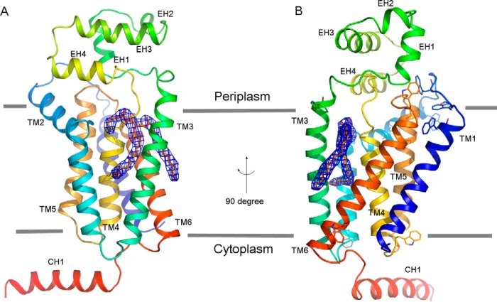
FIGURE 3.
Overall structure of the PgpB-PE complex. A, front view in the membrane plane. B, side view rotated in a 90° direction. The PgpB protein drawn as rainbow schematic contains a TM domain formed by TMs 1–6, an extracellular domain formed by EHs 1–4, and an intracellular helix CH1. PE lipid depicted as yellow sticks is superimposed with the 2Fo − Fc electron density map (blue meshes) contoured at 1.0 σ. The membrane boundaries (gray lines) are estimated from the positions of the multiple tryptophan residues (sticks) on both membrane surfaces.

