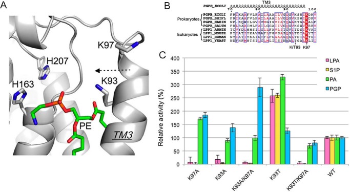
FIGURE 7.
Role of Lys-93 and Lys-97 on substrate selectivity. A, the conformation of Lys-93 and Lys-97 on TM3. PgpB is shown as a gray schematic. PE is depicted as green sticks. B, sequence alignment of TM3 from prokaryotic PgpBs (E. coli, Shigella flexneri, Haemophilus influenza, and Salmonella) and eukaryotic LPP1s (Arabidopsis thaliana, Mus musculus, Homo sapiens, and Saccharomyces cerevisiae). Conserved amino acids are white in red-filled rectangles. Identical residues are in red surrounded by blue lines. C, characterization of PgpB mutations at the Lys-93 and Lys-97 positions using four types of lipid phosphates. Activities were normalized to that of wild type.

