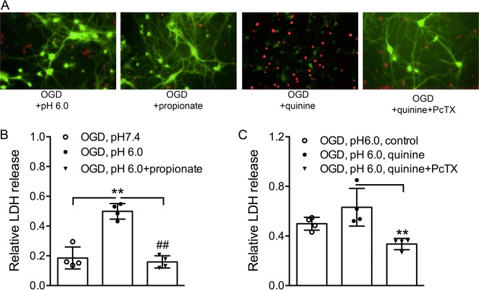
FIGURE 1.
Effect of quinine/propionate on OGD-induced injury of cultured mouse cortical neurons. A, fluorescein diacetate (green) and propidium iodide (red) staining of live and dead neurons taken at 6 h following 1 h of OGD treatment in the absence or presence of quinine or propionate shows that increasing intracellular pH (quinine) potentiates, whereas decreasing intracellular pH (propionate) inhibits OGD-induced neuronal injury. B, summary bar graph showing relative LDH release induced by OGD plus acidosis in the absence and presence of propionate. The LDH was increased by lowered extracellular pH, but reduced by propionate (10 mm) (one-way ANOVA, effect of treatment, F(2,9) = 43.55, p < 0.0001. Tukey's multiple comparisons test, **, p < 0.01 compared with pH 7.4; ##, p < 0.01 compared with pH 6.0 alone). C, summary bar graph showing that PcTX1 (10 nm) blocked enhancement of LDH release by quinine (1 mm) (one-way ANOVA, effect of treatment, F(2,9) = 9.5, p = 0.0061. Tukey's multiple comparisons test, **, p < 0.01 compared with quinine alone).

