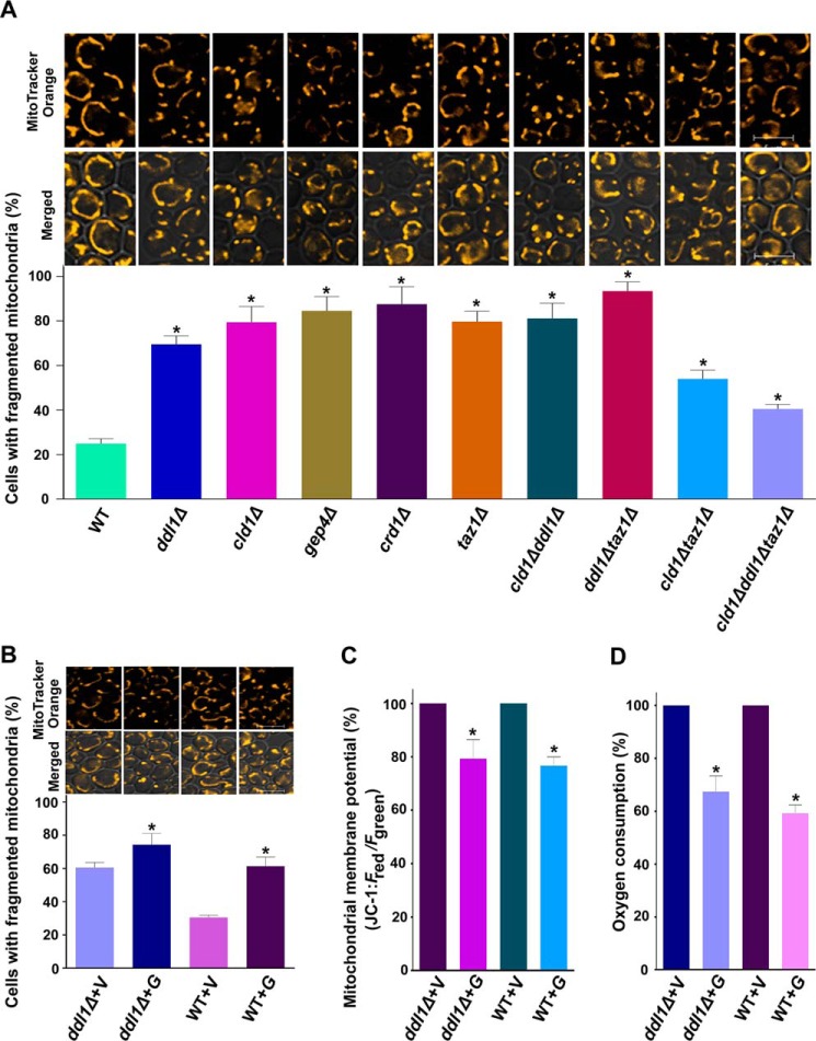
FIGURE 8.
Physiological significance of the DDL1 gene. A and B, the role of the DDL1 gene in mitochondrial morphology. Stationary-phase cells were stained with the mitochondrial dye MitoTracker Orange CMTMRos. The images were captured with a confocal microscope. C, study of the mitochondrial membrane potential. The cells were collected from the stationary phase and stained with the JC-1 dye. The ratio of Fred/Fgreen was used to determine the relative mitochondrial membrane potential. D, DDL1 overexpression and the oxygen consumption rate. The cells were collected from the stationary phase, and the oxygen consumption rate was analyzed. Bar, 5 μm. V, pYES2/NT B vector; G, pYES2/NT B-DDL1. To quantify mitochondrial fragmentation, at least 200 cells were scored in each background. The values are presented as the mean ± S.E. (n = 3). Significance was determined at *, p < 0.05.

