
Fig. 1.
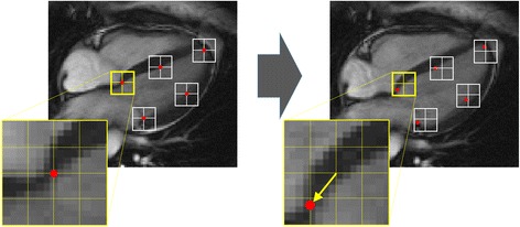
Basic principle of tissue tracking. Tracking the portions of tissue about a series of point (indicated in red on the pictures) is based on defining small square windows centered about such points on a first image (left picture) and searching the as-much-as-possible-similar grayscale pattern on the following image (right picture) in the vicinity of the original window. At the first step, shown here, the search windows have the same position in the pair of images and the feature that was at the center of the window of the first images is sought on the corresponding window on the second image (red dot) to provide an estimate of the local displacement. This procedure is usually repeated moving the second windows at the center of the new position and using a so-called coarse-to-fine approach, reducing progressively the window size. Large windows permit to recognize large gross displacements of the tissue, while the reduction of the window search area about the previous estimated targets allows to improve the accuracy and the locality of the estimation