
Fig. 2.
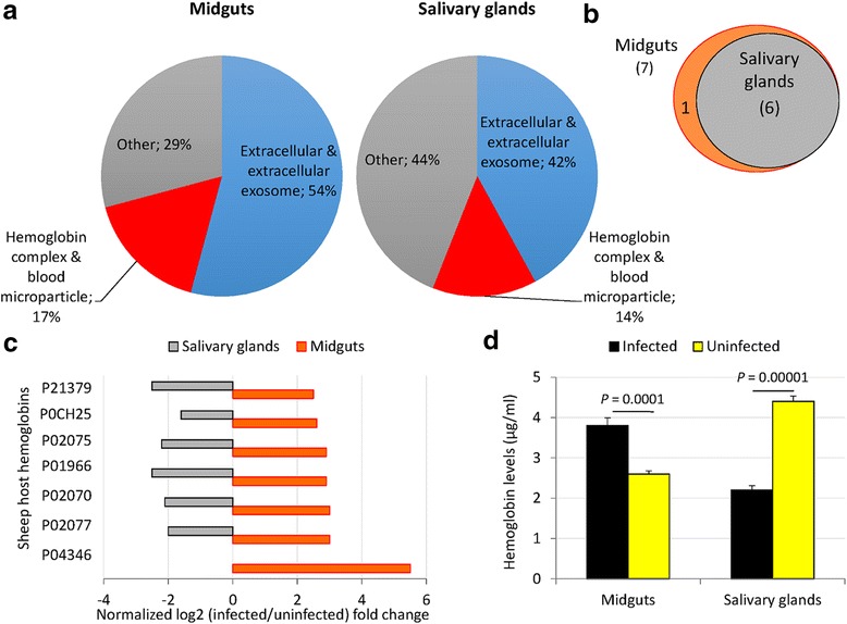
Sheep host hemoglobin levels vary in a tissue-specific manner in response to A. phagocytophilum infection in ticks. a Cell compartment classification of differentially represented sheep host proteins in infected female tick midguts and salivary glands. b Venn diagram of the sheep host hemoglobin differentially represented in infected vs uninfected tick tissues. c Differential host hemoglobin protein representation in response to A. phagocytophilum infection in tick midguts and salivary glands. d Hemoglobin levels in tick midguts and salivary glands from A. phagocytophilum-infected and uninfected ticks determined by ELISA in individual tick protein extracts, represented as the mean + standard deviation (SD) and compared between samples from infected and uninfected ticks by Student’s t-test with unequal variance (P < 0.05; 2 biological replicates)