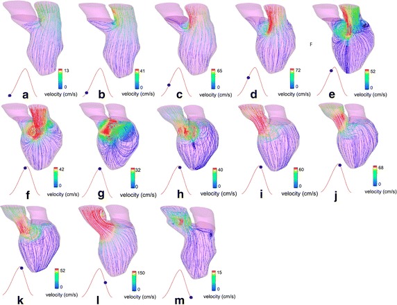
Fig. 7.

Flow patterns of an MI patient after surgery: Flow pattern during diastole (a–f) and systole (g–m), respectively. Strong vortices are formed during diastole in comparison to the pre-operative model (Fig. 6), which demonstrates the improvement in blood flow circulation after SVR. Improvement of the outflow jet direction through the aortic orifice demonstrates more efficient blood pumping after operation [13] (Adapted from [13], with permission from Wiley)
