Fig. 11.

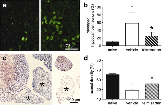
Telmisartan prevents irreversible neuronal damage and loss in the brains and spinal cords of NSV-infected mice. Fluoro-Jade C staining shows extensive labeling of hippocampal neurons in a mouse 7 days after NSV infection (right panel, a) compared to an uninfected control (left panel, a). Bar = 75 μm for both panels. Quantification of staining at this stage of infection as described in the “Methods” section shows that telmisartan (100 mg/kg/day) reduces damage to these neurons compared to animals treated with a vehicle control (b). In the lumbar spinal cord, silver staining shows prominent axonal loss in individual ventral nerve roots (each marked with an *) on day 7 post-infection (right panel, c) compared to an uninfected control (left panel, c). Bar = 100 μm for both panels. Quantification of axonal density in these ventral nerve roots as described in the “Methods” section shows that telmisartan treatment (100 mg/kg/day) reduces damage to lumbar motor neurons from which these axons arise compared to animals treated with a vehicle control (d). Student’s t test was used to analyze both the degree of cell damage in vehicle-treated versus naïve mice (†p < 0.0001 in both the hippocampus and spinal cord) as well as the degree to which drug treatment suppressed neuronal damage compared to those from vehicle-treated controls (*p = 0.0006 in the hippocampus; *p = 0.0005 in the spinal cord)
