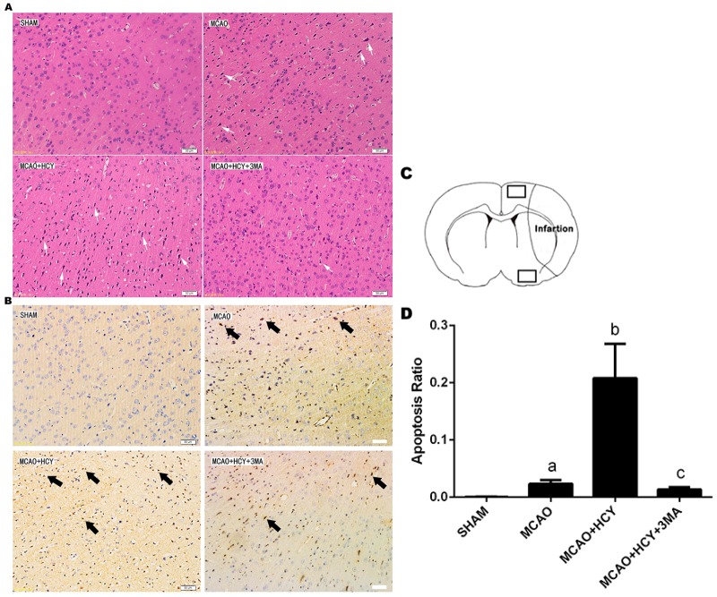
Figure 1.
The effect of Hcy on neuronal death in ipsilateral cortex penumbra after rat focal cerebral ischemia-reperfusion. (A) Histologic outcomes of HE staining; (B) Photomicrographs of the cortex penumbra of rat brains used for the TUNEL assay. The positive cells were stained in dark brown and are indicated by arrows; (C) Schematic showing examples of the areas (black squares) that were selected for counting of apoptotic cells in the cortex penumbra; (D) Quantification of apoptotic cells by the TUNEL assay. Apoptosis was expressed as the ratio of apoptotic cells to total cells. The data are expressed as ± s. a p < 0.05 vs. SHAM group; b p < 0.05 vs. MCAO group; c p < 0.05 vs. MCAO + HCY group. n = 3/group. Scale bars = 50 μm.

