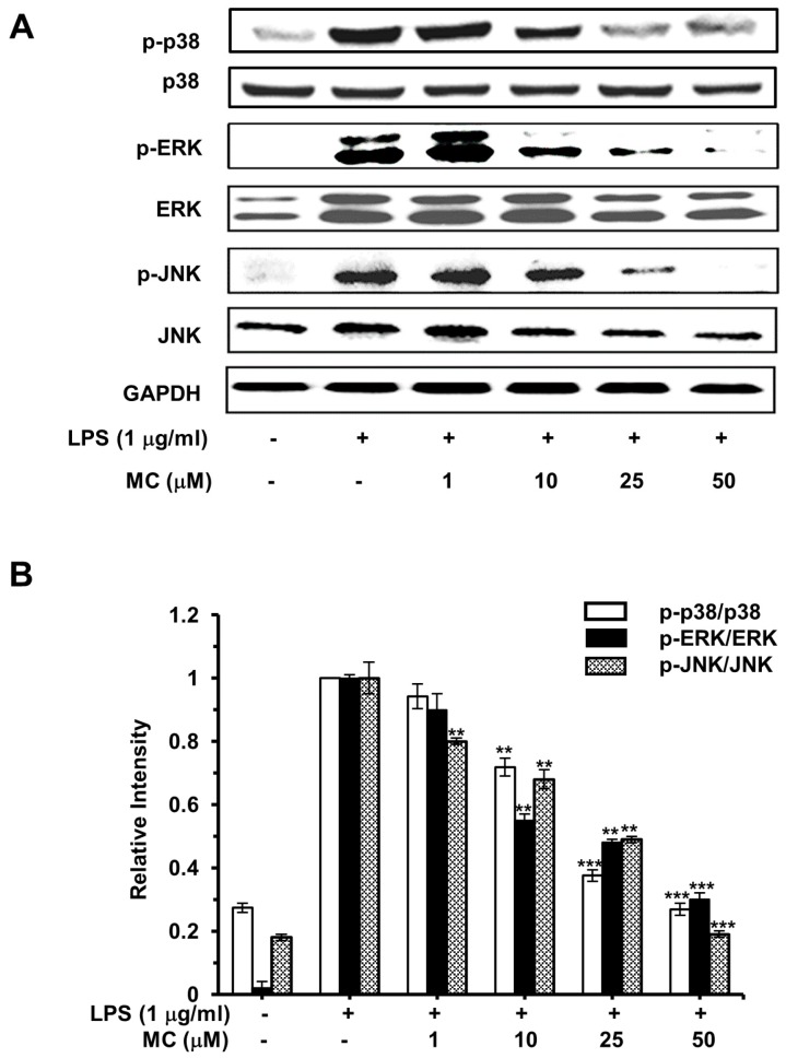
Figure 8.
Effects of MC on the phosphorylation of MAPKs. (A) Cells preincubated with 1, 10, 25 and 50 µM MC for 2 h were exposed to 1 µg/mL LPS for 30 min, for the detection of phosphorylated and total p38, ERK and JNK, cell lysates were extracted and measured by Western blotting analysis; (B) The ratio of the phosphorylation and the non-phosphorylation of MAPKs. The representative Western blotting bands from three independent experiments are shown here. Mean ± SEM, ** p < 0.01, *** p < 0.001.

