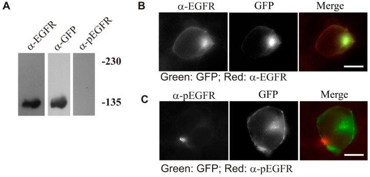
Figure 5.
Expression, phosphorylation and subcellular localization of ΔED-EGFR-GFP. 293T cells were transiently transfected with ΔED-EGFR-GFP. (A) The expression and phosphorylation of ΔED-EGFR-GFP. Following the transfection for 48 h, the cell were lysed and total cell lysates were used to determine the expression and phosphorylation of ΔED-EGFR-GFP by immunoblotting; (B) Subcellular distribution of ΔED-EGFR-GFP. Following the transfection for 48 h, the subcellular localization of ΔED-EGFR-GFP was revealed by the intrinsic GFP and by anti-EGFR antibody followed by TRITC-conjugated secondary antibody; (C) The phosphorylation of ΔED-EGFR-GFP. Following the transfection for 48 h, the phosphorylation of ΔED-EGFR-GFP was revealed by anti-pEGFR antibody followed by TRITC-conjugated secondary antibody. Size bar = 20 µm.

