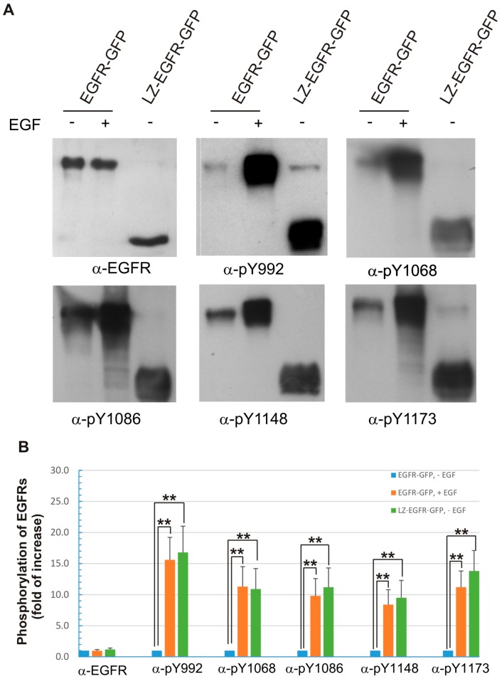
Figure 6.
Phosphorylation of the five major C-terminal tyrosine residues of EGFR-GFP and LZ-EGFR-GFP. (A) 293T cells were transiently transfected with EGFR-GFP or LZ-EGFR-GFP. Following serum starvation for 24 h, cells were treated with or without EGF. The cell lysates were subjected to immunoblotting analysis with rabbit anti-pEGFR (pY992), anti-pEGFR (pY1068), anti-pEGFR (pY1086), anti-pEGFR (pY1148) and anti-pEGFR (pY1173) antibodies; (B) Quantification of the data from (A). The band is quantitated by densitometry with image J software. The phosphorylation level of the control (EGFR-GFP, without EGF treatment) was set to 1 and the phosphorylation of the receptors under other conditions was expressed as the fold increase compared to control. Each value is the average of at least three independent experiments and the error bar is the standard error. **: p < 0.01.

