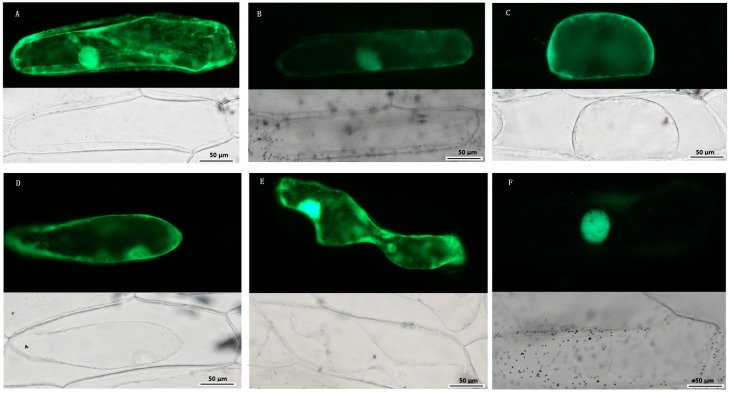
Figure 8.
Subcellular localization of TCS proteins. Green fluorescent protein (GFP)-fusion proteins were transiently expressed in onion epidermis cells under the control of the 35S promoter. After 16–18 h of incubation, GFP signal was detected with a green fluorescence microscope. Fluorescence (up) and bright-field images (down) of plasmolyzed empty vector pFGC: GFP transgenic cell (A); Fluorescence (up) and bright-field images (down) of 35S::SlHK8-GFP (B); 35S::SlHP2-GFP (C); 35S::SlHP3-GFP (D); 35S::SlRR1-GFP (E); and 35S::SlRR8-GFP (F) transgenic cell. Scale bar was presented in bottom right.

