Figure 1.
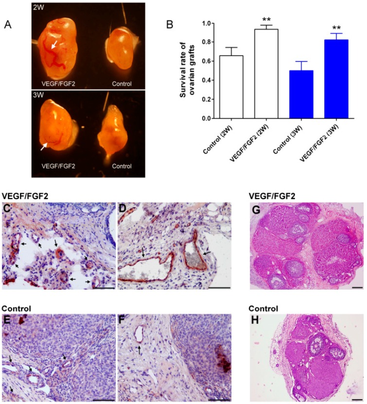
Morphology, survival rate, and blood vessels of the cryopreserved ovarian tissue after transplantation. (A) The morphologies of the grafted ovarian tissue. Left: VEGF/FGF2-treated tissue; right: untreated control tissue. An arrow shows the larger blood vessel; (B) The survival rate of the grafted ovarian tissue two and three weeks after transplantation. Survival rate is defined in the text. ** p < 0.01 compared with relative control group; (C–F) Immunohistochemical staining of vessels. The von Willebrand factor (vWF) protein, a marker of the endothelial cells of blood vessels, was stained red-brown (where arrow pointed); (G,H) Representative photos of H&E-stained ovarian sections show the morphology of ovarian grafts two weeks after transplantation. Scale bars: 100 µm.

