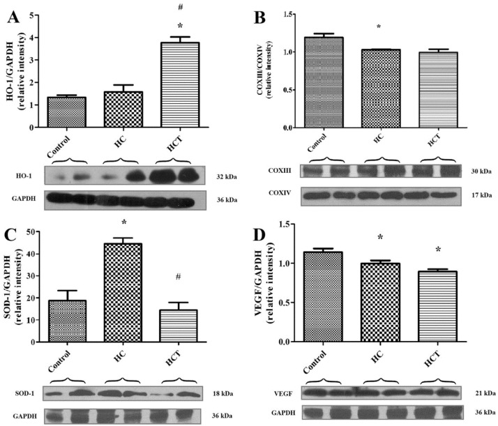
Figure 3.
Cardiac tissue biomarker expression: Western blot outcomes. The expression of HO-1 (A), COXIII (B), SOD-1 (C) and VEGF (D) protein in rabbit myocardial tissue was measured in homogenized left ventricular cardiac tissue samples drawn from three test groups (n = 6), defined as follows: I/R injured hearts from non-hypercholesterolemic animals fed with normal, non-cholesterol supplemented chow (control); I/R injured hearts from hypercholesterolemic animals fed with 2% cholesterol-supplemented chow (HC); and I/R-injured hearts harvested from hypercholesterolemic animals fed with 2% cholesterol and 2% wild garlic leaf lyophilisate-supplemented chow (HCT). GAPDH and COXIV expression levels were measured as reference proteins. Western blot analysis was conducted on each tissue homogenate in duplicate, and the signal intensity of the resulting bands corresponding to proteins of interest was measured using the Scion for Densitometry Image program, Alpha 4.0.2.3. The tissue content of each protein is shown in arbitrary units as the mean for each group of animal ± SEM. * p < 0.05 for comparison of the average expression levels of HO-1, COXIII, SOD-1 and VEGF in myocardium to the non-hypercholesterolemic group (control); # p < 0.05 for comparison to the hypercholesterolemic group (HC).

