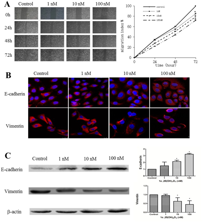
Figure 1.
1α,25(OH)2D3 inhibited the migration of human ovarian adenocarcinoma cell line SKOV-3 cells. (A) Left: representative pictures of the wound area obtained 24, 48 and 72 h after scratching. 100× magnification; Right: migration index (%) = [(the initialized width of the scratch) − (the final width of the scratch)]/(the initialized width of the scratch); (B) representative pictures of E-cadherin and Vimentin were captured by confocal laser scanner microscopy (CLSM) 24 h after being treated with 1α,25(OH)2D3. Nuclear DNA was visualized by 4′,6-diamidino-2-phenylindole (DAPI) staining. 200× magnification; (C) Left: Western blot analysis of the indicated proteins in SKOV-3 cells. β-actin served as a loading control; Right: the level of the indicated protein was quantified with gray value. The data represent the Mean ± SD. * p < 0.05 versus control.

