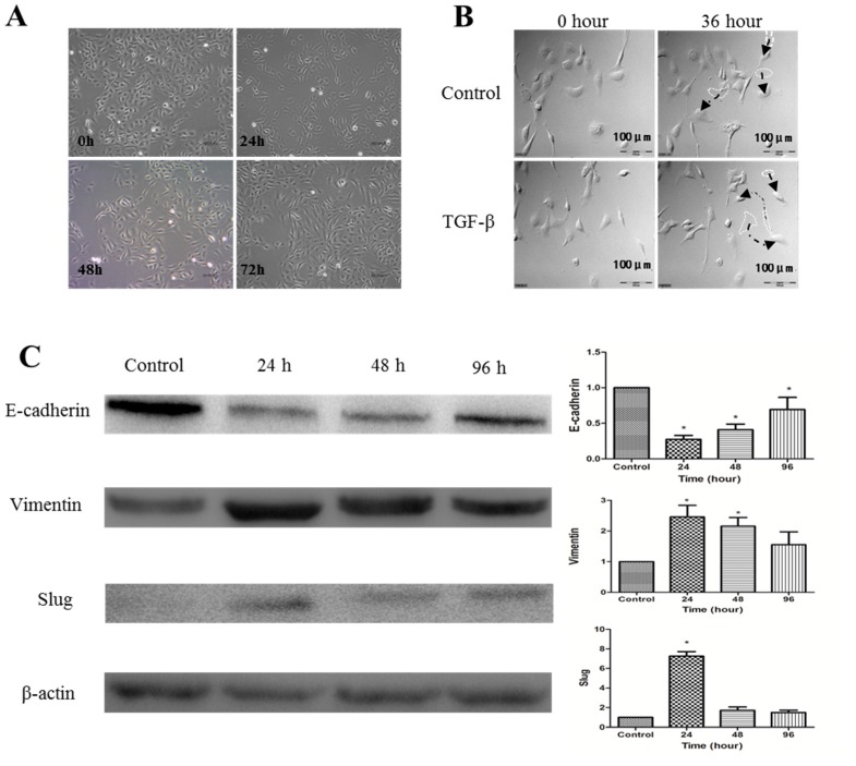
Figure 2.
Transforming growth factor-β1 (TGF-β1) induces EMT of SKOV-3 cells. (A) SKOV-3 cells were exposed to 10 ng/mL of TGF-β1. Compared to the group of control, TGF-β1-treated SKOV-3 cells lost their cobblestone shape and adopted a fibroblast-like, spindle-shaped morphology. Morphology photographs were taken at 24, 48, and 72 h (magnification of 400×); (B) analyses with a Live Cell Imaging System showed that the movement distance increased further after SKOV-3 cells were treated with TGF-β1 for 36 h than control cells (Arrows refer to the movement track of SKOV-3 cells); (C) Left: Western blot analysis of the indicated proteins in SKOV-3 cells. β-actin served as a loading control; Right: the level of the indicated protein was quantified with gray value. The data represent the Mean ± SD. * p < 0.05 versus control.

