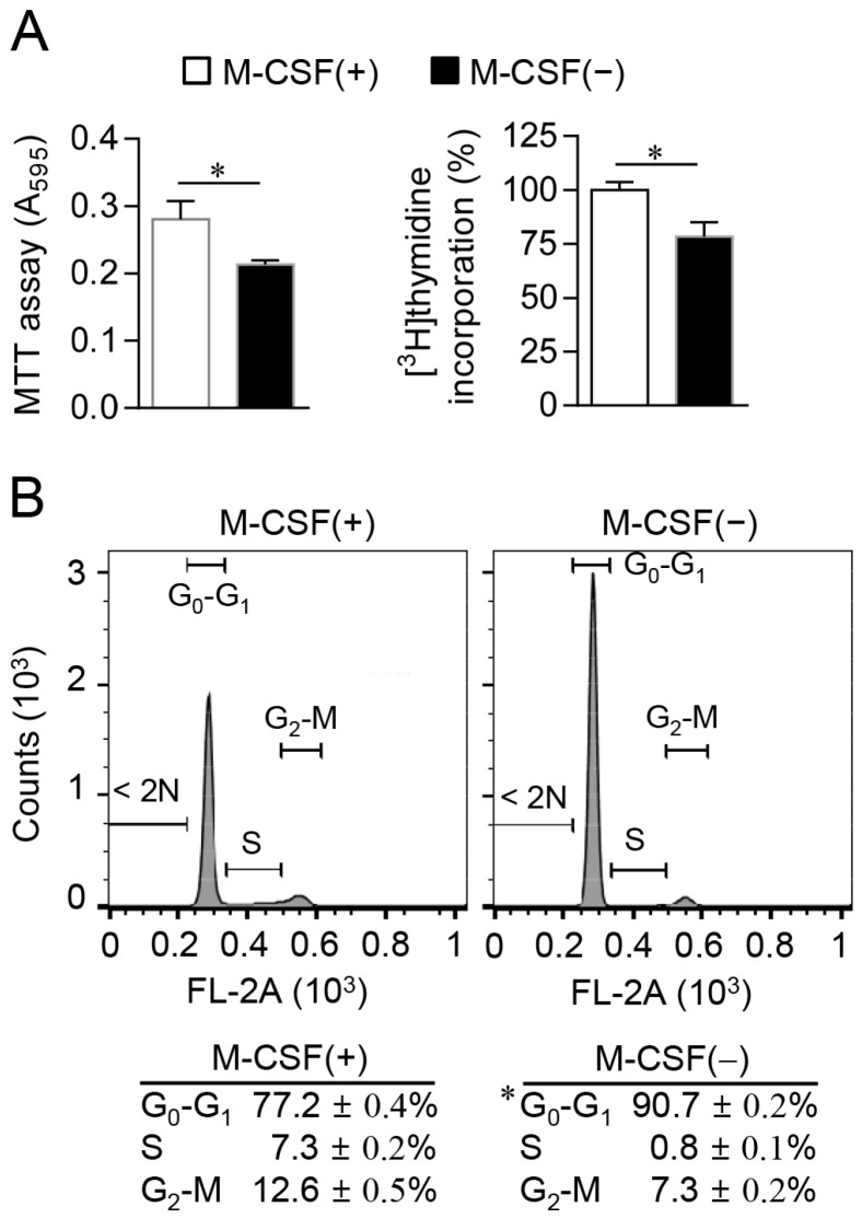
Figure 2.
Induction of G0–G1 cell cycle arrest in osteoclast progenitors by M-CSF deprivation. (A) Cells were cultured in the absence or presence of M-CSF for 12 h, after which cell proliferation was determined with the MTT assay (left panel) or by measurement of [3H]thymidine incorporation (right panel); (B) Cells cultured as in (A) were stained with propidium iodide and subjected to cell cycle analysis by flow cytometry. Data are means ± SD for a representative experiment run in triplicate. * p < 0.01 (Student’s t test).

