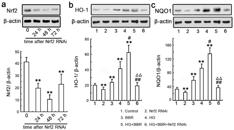
Figure 7.
siRNA knockdown of Nrf2 abrogates BBR-induced NQO1 and HO-1 expression. (a) NRK-52E cells were transfected with Nrf2-siRNA and western blot analysis was performed with an antibody against Nrf2 was performed at various time-points following transfection (24, 48 and 72 h). Relative Nrf2 expression levels were calculated and normalized to the loading control. Corresponding protein levels were assessed using densitometry and expressed in relative intensities. All results were obtained from three independent experiments. Values are expressed as the mean ± SEM (n = 6; ** p < 0.01, vs. control); (b,c) The cells were divided into six groups as description in this paper. Western blotting was performed with an antibody against NQO1 and HO-1. Relative NQO1 and HO-1 expression levels were calculated and normalized to the loading control. Corresponding protein levels were assessed using densitometry and were expressed in relative intensities. All results were obtained from three independent experiments. Values are expressed as the mean ± SEM (n = 6) (** p < 0.01 vs. control; ## p < 0.01 vs. HG; # p < 0.05 vs. HG; ΔΔ p < 0.01 vs. HG + BBR).

