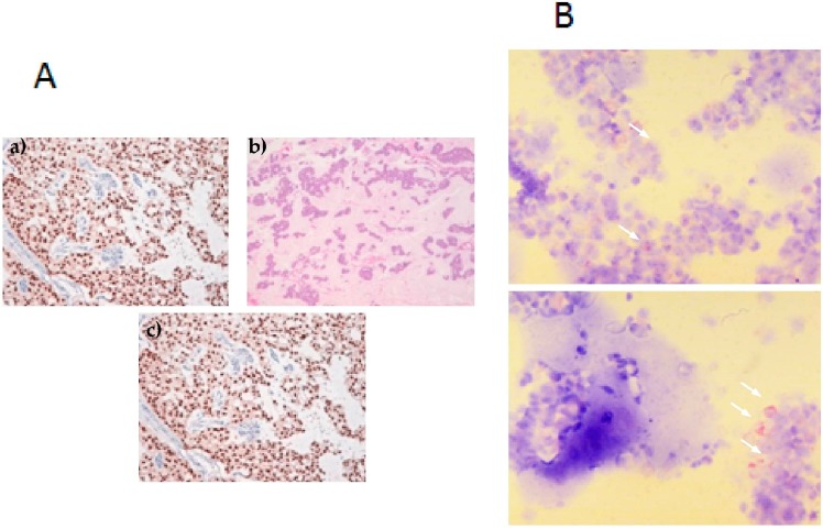
Figure 1.
Breast cancer bone metastasis. (A): (a) Histological hematoxylin and eosin (H & E) staining of bone metastasis (BM) primary culture (5× magnification). Mucinous areas show low cellularity; (b) H & E staining of BM primary culture (20× magnification). Monomorphic cells with round nucleus and nucleolus are seen in nests with a minor mucus quantity; (c) ER staining showing positivity; (B): Cytospin of BM primary cells. White arrows show pancytokeratin-positive cells.

