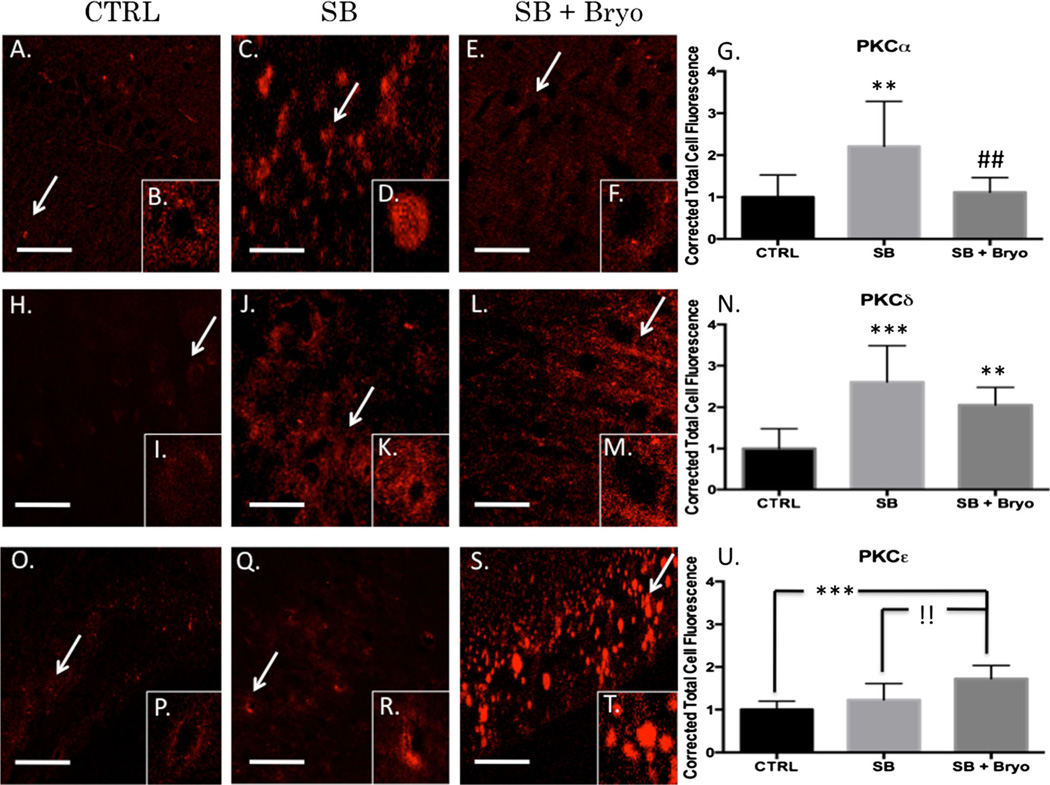
Fig. 2.
Bryostatin-1 decreases PKCα and increases PKCε 24 h after blast exposure. Our data show that bryostatin-1 has a profound effect after blast traumatic brain injury using fluorescent IHC. Scale bar=100 µm in left prefrontal cortex. PKCα control (a) with inlay (b) compared to single blast exposure (c) with inlay (d), and single blast exposure + bryostatin-1 (e) with inlay (f) showed significant difference between groups. Post-hoc comparison between control and single blast (**p<0.01) and between single blast and single blast + bryostatin-1 (##p<0.01) as depicted in bar graph (g). PKCγ control (h) with inlay (i) compared to single blast exposure (j) with inlay (k), and single blast exposure + bryostatin-1 (l) with inlay (m) showed significant difference between groups. Post-hoc comparison between control and single blast (***p<0.001) and between control and single blast + bryostatin-1 (**p<0.01) as depicted in bar graph (n). PKCε control (o) with inlay (p) compared to single blast exposure (q) with inlay (r), and single blast exposure + bryostatin-1 (s) with inlay (t) showed a significant difference between groups. Post-hoc comparison between control and single blast + bryostatin-1 (***p<0.001) and between single blast and single blast + bryostatin-1 (!!p<0.01) as depicted in bar graph (u)

