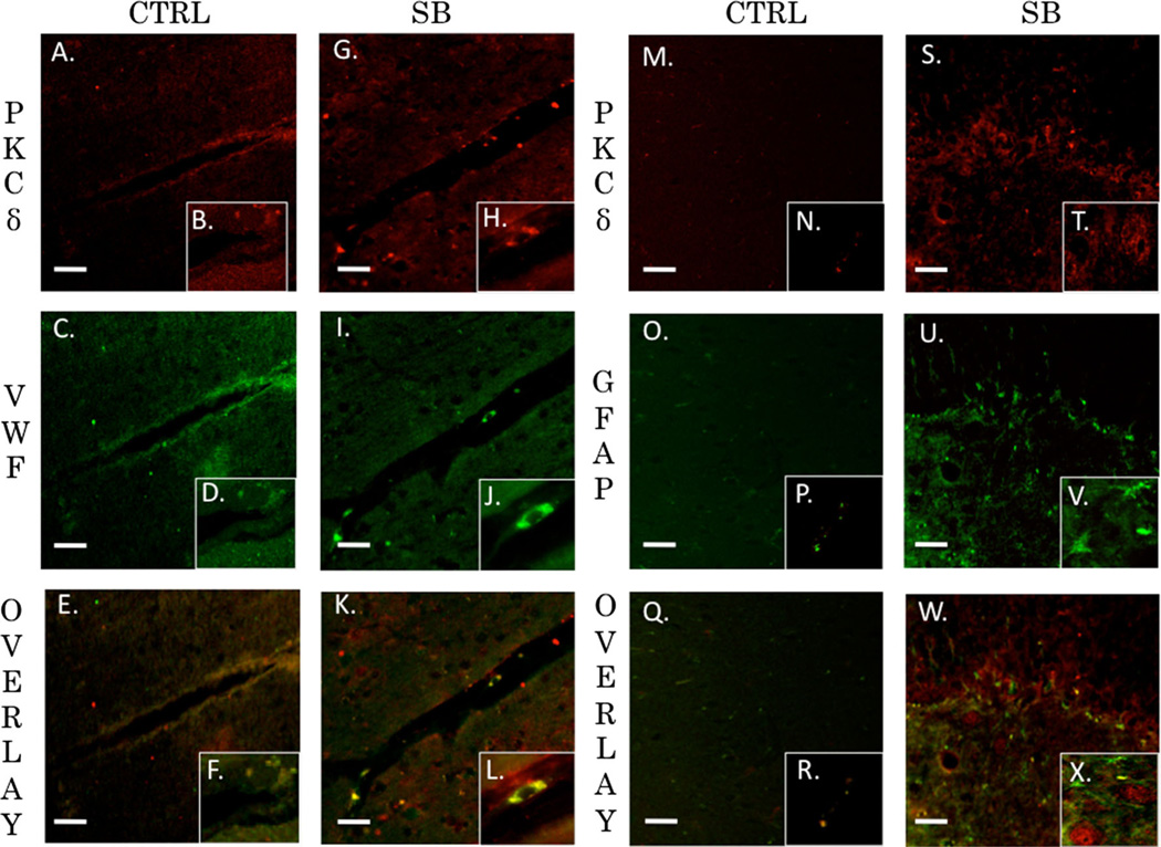
Fig. 9.
PKCδ co-localized with endothelial cells but not astrocytes. PKCδ plays an important role in mediating vascular tone. Its role in regulation of tight junction proteins is not completely understood. Fluorescent IHC red staining for PKCδ, green staining for VWF (endothelial) or GFAP (astocyte), and yellow is overlay. Scale bar=20 µm in left prefrontal cortex. PKCδ (a) with inlay (b) and VWF (c) with inlay (d) have a moderate overlay with a Pearson’s coefficient of r=0.61 seen in (e) with inlay (f) for control animals. PKCδ (g) with inlay (h) and VWF (i) with inlay (j) have a strong overlay with an overlap coefficient of r=0.88 seen in (k) with inlay (l) 24 h post blast exposure. PKCδ (m) with inlay (n) and GFAP (o) with inlay (p) have a very weak overlay with a Pearson’s coefficient of r=0.029 seen in (q) with inlay (r) for control animals. PKCδ (s) with inlay (t) and GFAP (u) with inlay (v) have a moderate overlay with an overlap coefficient of r=0.449 seen in (w) with inlay (x) 24 h post blast exposure

