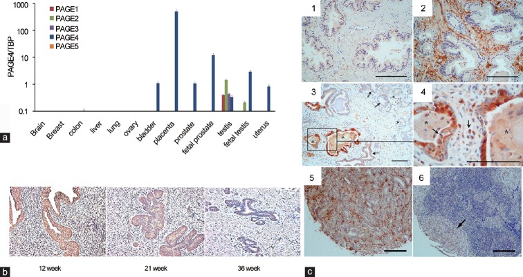
Figure 1.
PAGE expression at the mRNA and protein levels. (a) Expression of mRNAs encoding PAGE1-5 in different normal human tissue samples obtained from healthy donors. (b) Immunohistochemistry analysis of PAGE4 in the human fetal prostate. Samples were stained for PAGE4 at gestational weeks 12, 21 and 36. (c) Immunohistochemistry analysis of PAGE4 in prostate cancer. (1) Negative staining in the normal prostate. (2) Intense staining shown in the stromal tissue in BPH. (3) Intense staining shown in cancer adjacent “normal” glands (asterisk) associated with inflammation but only moderate staining in the cancer cells (arrowhead). (4) High-power view of boxed area in (3). (5) PAGE4 protein expression in organ-confined prostate cancer. (6) Loss of PAGE4 protein expression in metastatic prostate cancer. Asterisk: PIA lesions; arrows indicate inflammatory cells. Scale bars in all panels: 100 μm. Data presented are reproduced with permission from original publications by the authors.

