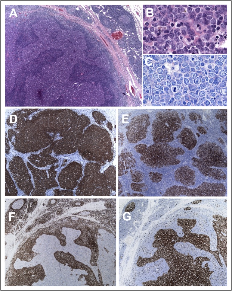
Figure 1.
Histologic features of nodal PTFL. (A) The nodal architecture is effaced by ill-defined, coalescent follicles. A starry sky pattern is evident. The mantle zone is attenuated. Note a rim of residual normal nodal tissue with residual GCs (hematoxylin-and-eosin [H&E] stain; original magnification, ×25). (B) Cytologic features of the tumor cells within the abnormal follicles. The infiltrate is composed of medium-sized cells with blastic chromatin and inconspicuous nucleoli (H&E stain; original magnification, ×400). (C) Giemsa stain highlights the presence of medium-sized blastoid cells with few scattered centroblasts (original magnification, ×400). (D) CD20 shows the abnormal follicles, but CD20+ cells do not extend to the interfollicular region. (E) The follicles are strongly CD10+. (F) The follicular cells are BCL2−. (G) MIB1 stain demonstrates the high proliferation within the follicles without polarization (D-G, immunoperoxidase; original magnification, ×25).

